The First Annual “Stalling Summit”
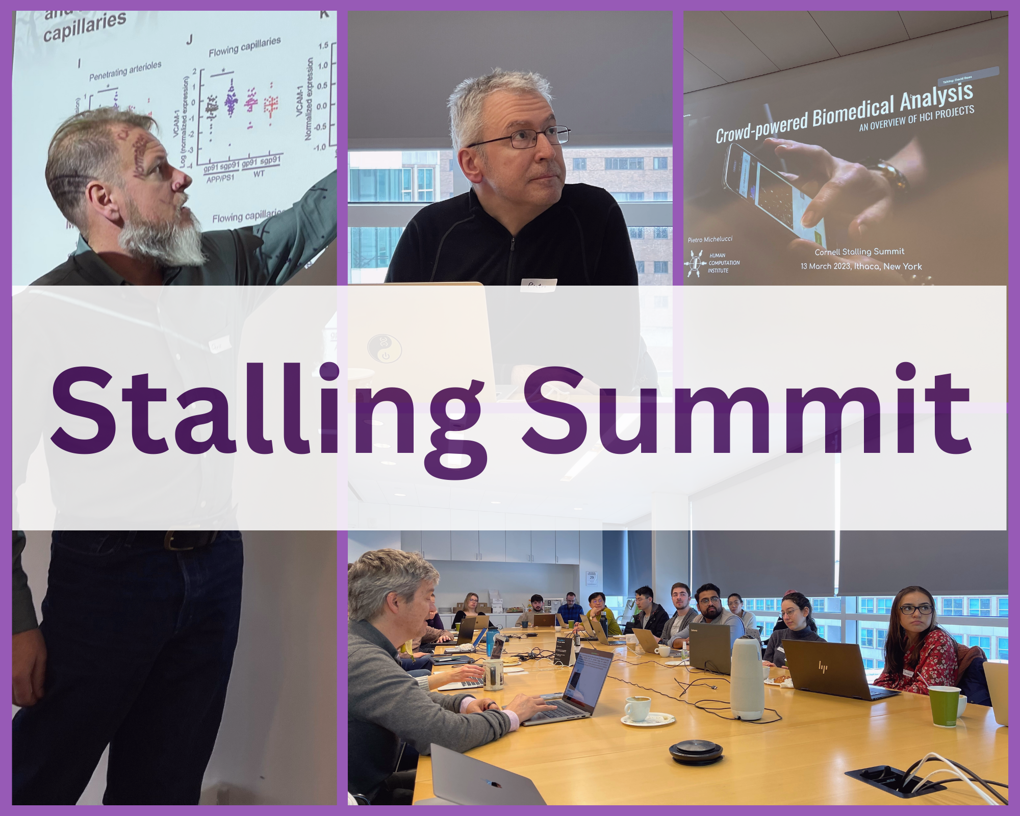
What happens when world class Alzheimer’s researchers and stroke researchers put their heads together to help each other? Among other things, they turn to Stall Catchers with some new requests for help from our loyal community of catchers!
As it turns out both Alzheimer’s and stroke seems to be asking related questions related to brain blood flow and the ability to deliver oxygen to neurons, the cells in our brains that process information when we are thinking. In the Stall Catchers Alzheimer’s work so far, we have been trying to understand the role of capillary stalling in both cognitive symptoms and the progression of Alzheimer’s disease, and we’ve already learned a lot (link to research findings blog post).
In stroke research, we are trying to understand how restoring blood flow after a brain clot is related to getting memories and other brain functions back, like motor control. In both cases, to answer these research questions, we use “mouse models” of Alzheimer’s disease and stroke, and then we use special imaging techniques to see what’s going in with blood flow in the tiny capillary vessels.
Our newest collaborators at Boston University, led by Professor David Boas, are bringing some exciting new imaging techniques that add to our toolbox for investigating blood flow. In particular, they use a special optical imaging technique, which is going to be a game-changer for us. In Stall Catchers, we use two-photon excitation microscopy (2PEM) to create a vessel movie that shows blood flow over time.
With our current "Gaussian beam" version of 2PEM, we can only image one layer of brain tissue at a time, so as time passes in the vessel movie, we are looking through successively deeper layers of brain tissue with a very high frame rate. This allows us to see changes from moment to moment, but only one layer at a time.
In comparison, another approach called optical coherence tomography (OTC) would allow us to see through all layers of brain tissue simultaneously. The problem is OTC is very slow, so the time gap between each frame makes it hard to see whether or not red blood cells are moving through vessels or not.

Enter the “magical” Bessel beam technique! Bessel beam is another form of 2PEM imaging, which collapses all layers of blood vessels into a single imaging layer (just like OTC) and can do it much faster. So it combines the best features of the Gaussian beam 2PEM we are currently using for Stall Catchers and OTC. This combination makes it potentially easier to see the hemodynamics (blood flow) of a capillary throughout the entire vessel movie.
What does this mean for the future of Stall Catchers? We will likely start using Bessel beam data for our Alzheimer’s research AND start to analyze new data sets that help our Bostonian stroke researchers.
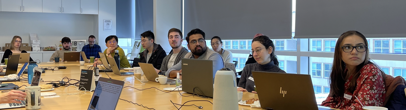
The good news is that the relationship between the two research programs means that any findings from one lab help advance the research in the other lab. In fact, there will be monthly collaboration meetings with both our Cornell-based collaborators and Boston University collaborators, to ensure the best opportunities for crosstalk and speeding up research in both places!
A big welcome to the Boston University crew of researchers and looking forward to writing this next chapter of Stall Catchers with all of you.